Chapter 4
Foundations of biology
Life, cells, and how biologists work
Human Biology for the Medical Maker
Why this chapter exists
- Modern medicine is applied cell biology — insulin, tumour sequencing, mRNA vaccines, CAR-T
- The laboratory is technology-dense — microscopes, incubators, sequencers, robots, software
- Cells set physical limits — diffusion, growth, contamination, biofilm
Enough to read a methods section, follow a lab conversation, and know when a question needs a biologist.
What counts as life
| Property | Engineering reading |
|---|
| Organisation | A hierarchy of modules |
| Metabolism | An open system far from equilibrium |
| Homeostasis | Feedback control |
| Growth and development | A build process driven by stored instructions |
| Reproduction with heredity | Self-replication with an error rate |
| Response to environment | Sensors, actuators, signalling |
| Evolution | An optimiser with no foresight |
Viruses sit on the boundary — they are not cells, so antibiotics do nothing to them.
The two organising ideas
Cell theory
- All living things are made of cells
- The cell is the basic unit of life
- Cells arise only from existing cells — so contamination must come from somewhere
Evolution by natural selection
- Bodies are not designed, and it shows
- Variation is the normal state
- Microbes evolve fast: antibiotic resistance in real time
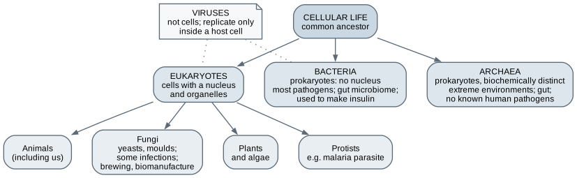
The tree of life
- Bacteria and archaea: prokaryotes, no nucleus
- Eukaryotes: nucleus and organelles — animals, fungi, plants, protists
- Viruses infect every domain
Prokaryotic versus eukaryotic cells
| Feature | Bacterium | Human cell |
|---|
| Size | 1 to 2 micrometres | 10 to 30 micrometres |
| DNA | One circular chromosome, plus plasmids | Linear chromosomes in a nucleus |
| Ribosomes | Smaller (70S) | Larger (80S) |
| Cell wall | Usually, peptidoglycan | None in animals |
| Division | About 20 to 30 minutes at best | About 24 hours in culture |
The differences are what antibiotics target. Selectivity needs a difference.
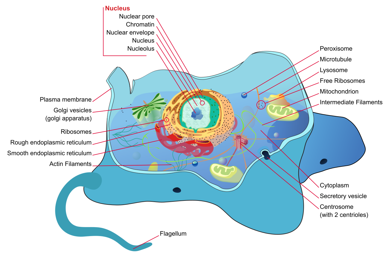
The eukaryotic cell
- Nucleus: about 2 metres of DNA in 5 to 10 micrometres
- Ribosomes, rough ER, Golgi: the protein production line
- Mitochondria: most of the ATP; their own DNA
- Lysosomes: digestion
- Cytoskeleton: shape, transport, and sensing load
Scale, and the limit of the light microscope
| Structure | Typical size |
|---|
| DNA helix, membrane, ribosome | 2 nm, 7 to 8 nm, about 25 nm |
| Virus | 20 to 300 nm |
| Light microscope limit | about 0.2 micrometres |
| Bacterium | 1 to 2 micrometres |
| Red blood cell; typical human cell | 7 to 8; 10 to 30 micrometres |
| Human egg; neuron axon | about 0.1 mm; up to about 1 m long |
Everything above the microscope limit row is invisible to light microscopy.
From DNA to protein: the central dogma
- DNA — the stored source; copied before division
- Transcription — a gene copied into pre-mRNA
- Processing — introns spliced out; cap and tail added
- Translation — ribosomes read codons and link amino acids
- Folding and modification — in the ER and Golgi, into a working protein
Regulation acts at every step.
DNA and its replication
The genome
- A pairs with T, G with C — each strand templates the other
- About 3.1 billion base pairs; about 20,000 genes
- Protein-coding sequence is only 1 to 2 percent
Copying it
- Tens of thousands of origins in parallel
- Proofreading plus repair: about 1 error per billion bases
- Telomeres cap and shorten the ends
- Radiotherapy works partly by overwhelming repair
Transcription, splicing, and the genetic code
- RNA polymerase copies a gene into mRNA
- Introns cut out, exons joined; alternative splicing makes variants
- Codons: 64 triplets, 61 amino acids, 3 stops; AUG starts
- Redundant: many single-base changes are silent
- Nearly universal: bacteria can make human insulin
The software analogy — and where it breaks
The analogy
- DNA is source code in storage
- mRNA is a checked-out working copy
- The ribosome is an interpreter
- Regulation is configuration
Where it breaks
- Massively parallel, rates set by chemistry
- Bugs are partial — 30 percent less function, not a crash
- Configuration is heritable: epigenetics
The cell cycle, division, and death
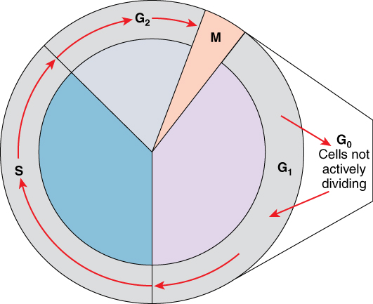
- G1 growth, S replication, G2 checking, M mitosis
- G0: neurons and heart muscle rest for life
- Checkpoints, p53: cancer is their failure
- Chemotherapy hits fast-dividing hair, gut, marrow
- Meiosis halves chromosomes and shuffles them
Specialisation and stem cells
| Type | Potential | Example |
|---|
| Totipotent | Every cell type including placenta | Fertilised egg |
| Pluripotent | Every body cell type | Embryonic stem cells; iPSCs from skin or blood |
| Multipotent | A family of types | Blood-forming stem cells; bone marrow transplant |
| Unipotent | One type | Skin and gut lining renewal |
"Stem cell therapy" is also an unregulated market. Ask: approved therapy, or registered trial?
Microbes at the cell level
Bacteria
- Gram-positive thick wall; Gram-negative outer membrane
- Spores survive heat and disinfectants
- Plasmids carry resistance between species
- Growth: lag, exponential, stationary, death
Viruses and biofilms
- Genome in a coat; 20 to 300 nanometres
- Attach, hijack, assemble, leave
- Biofilm: hundreds of times more tolerant of antibiotics
- A surface that can be colonised but not cleaned is the design problem
The immune system at the cell level
| Cell | Role |
|---|
| Neutrophils | First responders; engulf bacteria |
| Macrophages | Clear debris; wall off implants — the foreign body response |
| Dendritic cells | Present fragments to T cells; vaccine targets |
| T cells | Coordinate and kill; CAR-T re-engineers them |
| B cells | Make antibodies — basis of immunoassays and antibody drugs |
| Memory cells | Faster second response — why vaccines work |
Why tissues need blood vessels: diffusion time
| Distance | Time for oxygen | Meaning |
|---|
| 10 micrometres | About 25 milliseconds | Instantaneous |
| 100 micrometres | About 2.5 seconds | Still adequate |
| 1 millimetre | About 4 minutes | Too slow for living tissue |
| 1 centimetre | About 7 hours | Impossible |
t ≈ x² / 2D. Most cells sit within 100 to 200 micrometres of a capillary.
Exponential growth, and the genome as data
Growth
- Bacteria every 20 minutes: a billion cells in 10 hours
- Cell culture daily: a million to a billion in 10 days
- PCR: 30 cycles ideally 2³⁰; at 90 percent, five times less
Information
- 2 bits per base: about 775 MB per genome
- People differ at about 1 base in 1,000
- Copy errors 10⁻⁹ per base — poor for storage, essential for evolution
Seeing small things, and spinning things down
Resolution
- d ≈ λ / 2NA: about 200 nanometres for visible light
- Super-resolution: tens of nanometres
- Electron and cryo-EM: near-atomic
- Fluorescence: more light, more bleaching and damage
Centrifuges
- RCF ≈ 1.118 × 10⁻⁵ × r × rpm²
- 3,000 rpm at 10 cm ≈ 1,000 g
- A protocol in rpm does not transfer; one in g does
The branches of biology and their real-world uses
| Branch | Real-world use |
|---|
| Genetics and genomics | Newborn screening, rare disease diagnosis, tumour sequencing |
| Microbiology and virology | Hospital labs, infection control, sterility testing |
| Immunology | Vaccines, monoclonal antibodies, immunoassays |
| Biomanufacturing | Recombinant insulin, antibodies, mRNA vaccines |
| Cell and gene therapy | CAR-T; first CRISPR therapy approved 2023 |
| Tissue engineering and organoids | Skin substitutes, organ-on-chip drug testing |
| Bioinformatics | The most direct entry for a software-literate engineer |
The laboratory workflow
- Sample — blood, swab, tissue, cells
- Grow — incubators, culture, bioreactors
- Separate and prepare — centrifuge, pipettes, extraction, gels
- Measure — microscopes, plate readers, flow cytometry, PCR, sequencers
- Analyse — software, statistics, databases
- Record and store — notebooks, LIMS, freezers, biobanks
Tools for seeing and for growing
Seeing
- Brightfield: stained tissue and blood films
- Phase contrast: living unstained cells
- Fluorescence and confocal: labelled proteins, 3D
- Light-sheet; electron microscopy; cryo-EM
Growing
- Biological safety cabinet — not a fume hood
- CO₂ incubator: 37 °C, 5 percent CO₂, humidity
- Culture media and treated surfaces
- Bioreactors: oxygen and mixing without shear
Tools for preparing and measuring
Separating and preparing
- Micropipettes: the most-used instrument
- Liquid-handling robots
- Centrifuges and ultracentrifuges
- Nucleic acid extraction; gel electrophoresis
Measuring and counting
- Plate readers: ELISA, viability, screening
- Flow cytometers: thousands of cells per second
- Western blot, ELISA, mass spectrometry
- Patch clamp for ion channels
Tools for reading and editing genes
| Tool | What it does |
|---|
| PCR and qPCR | Amplify a sequence; infection diagnosis, genetic tests |
| Sanger sequencing | One fragment, up to about 1,000 bases, very accurate |
| Short-read sequencing | Billions of reads; a genome for hundreds of dollars in reagents |
| Long-read and nanopore | Tens of thousands of bases; pocket-sized devices |
| CRISPR | A guide RNA directs a cut at a chosen sequence |
Storing, records, and software
Storage and records
- Freezers to minus 80; liquid nitrogen at minus 196
- Freezer alarms: a classic monitoring problem
- Biobanks, LIMS, ELN
Software
- NCBI, Ensembl, UniProt, BLAST
- Biopython, Bioconductor, Galaxy
- Fiji, CellProfiler, napari: image analysis
- AlphaFold, PyMOL, ChimeraX
Biosafety levels
| Level | Examples | Key controls |
|---|
| BSL-1 | Lab strains of E. coli, baker's yeast | Good practice; open bench |
| BSL-2 | Staphylococcus, Salmonella, clinical samples | Safety cabinets, PPE, restricted access |
| BSL-3 | Tuberculosis, some respiratory viruses | Negative pressure, airlocks, respirators |
| BSL-4 | Ebola, Marburg | Suits, isolated buildings |
Do not culture microorganisms or human cells outside a properly supervised laboratory.
Where a technologist contributes
- Open-source microscopes: flexure stages, optics, camera, software
- Temperature control: PID, logging, alarms, enclosures
- Labware holders and adaptors: machined, moulded, or printed in resistant material
- Imaging enclosures; teaching bioreactors; microfluidics
- Image analysis on public data — no lab needed
Open hardware in a clinical lab is still a validation question. Build accessories, not rotors or power supplies.
Worked example: twelve gel imagers for a teaching lab
Questions first
- Blue-light stains only — no UV hazard
- Wipeable stage; check the lab's disinfectant
- Phones or a fixed camera?
- Never connected to the gel tank
Then method and acceptance
- Sheet metal or acrylic box, LED array, amber filter
- Prototype once, then batch with jigs
- Accept: 5 ng band visible, even light over 10 cm
- Price twelve commercial units first
Workshop exercise and exit criteria
- Look at onion and yeast cells; measure them
- Extract strawberry DNA with kitchen materials
- Measure diffusion in gelatin; plot against root time
- Watch yeast grow exponentially
- Read the insulin gene in NCBI; count nuclei in Fiji
What this chapter does not establish
- Knowing what an instrument measures is not knowing how to run it
- No clinical samples, no cultures, outside a lab's biosafety framework
- A home-built instrument is not a validated instrument
- Cell biology knowledge is not clinical knowledge
- The field moves fast — check current numbers
Next — Tissues, skin, and contact with an object.