Chapter 8
The head and neck: eye, ear, nose, throat, and mouth The special senses, the airway–food crossing, and the optics and acoustics behind labels and alarms
Human Biology for the Medical Maker
Welcome to chapter eight, the head and neck. Chapter seven treated seeing and hearing as capabilities that a design must not depend on alone. It never opened the organs that provide them. This chapter does. We will go through the eye, the ear, the nose and sinuses, the throat and the mouth, and then do the optics and acoustics that let you check a label or an alarm against human limits before you build anything. Why this chapter exists Four special senses, the airway, the food passage, the voice, in a shoebox Almost everything close to the surface; almost nothing padded A place devices sit: glasses, hearing aids, tubing, masks, straps The reason an interface works or fails: a label is optics, a beep is acoustics Simulator and training territory The airway and the food passage cross. Anything in a mouth is an airway question.
The head and neck are the densest region of the body for a designer. In a volume smaller than a shoebox sit four of the five special senses, the only route for air into the lungs, the only route for food into the gut, the place where those two routes cross, the voice and the teeth. Almost everything is close to the surface and almost nothing is padded. You will meet this region as a place devices sit, as the reason an interface works or fails, and as simulator territory. And one rule belongs at the start: the airway and the food passage cross. Anything that goes into the mouth is, until shown otherwise, something that can go into the lungs. The special senses and their nerves Sense Organ Receptor and stimulus Cranial nerve Vision Eye Rods and cones; light, 400 to 700 nm II Hearing Cochlea Hair cells; fluid vibration VIII Balance Vestibular apparatus Hair cells; rotation, acceleration, gravity VIII Smell Olfactory epithelium Airborne molecules I Taste Taste buds Dissolved molecules VII, IX, X
Corneal blink: sensation in on nerve V, command out on VII — any link can fail.
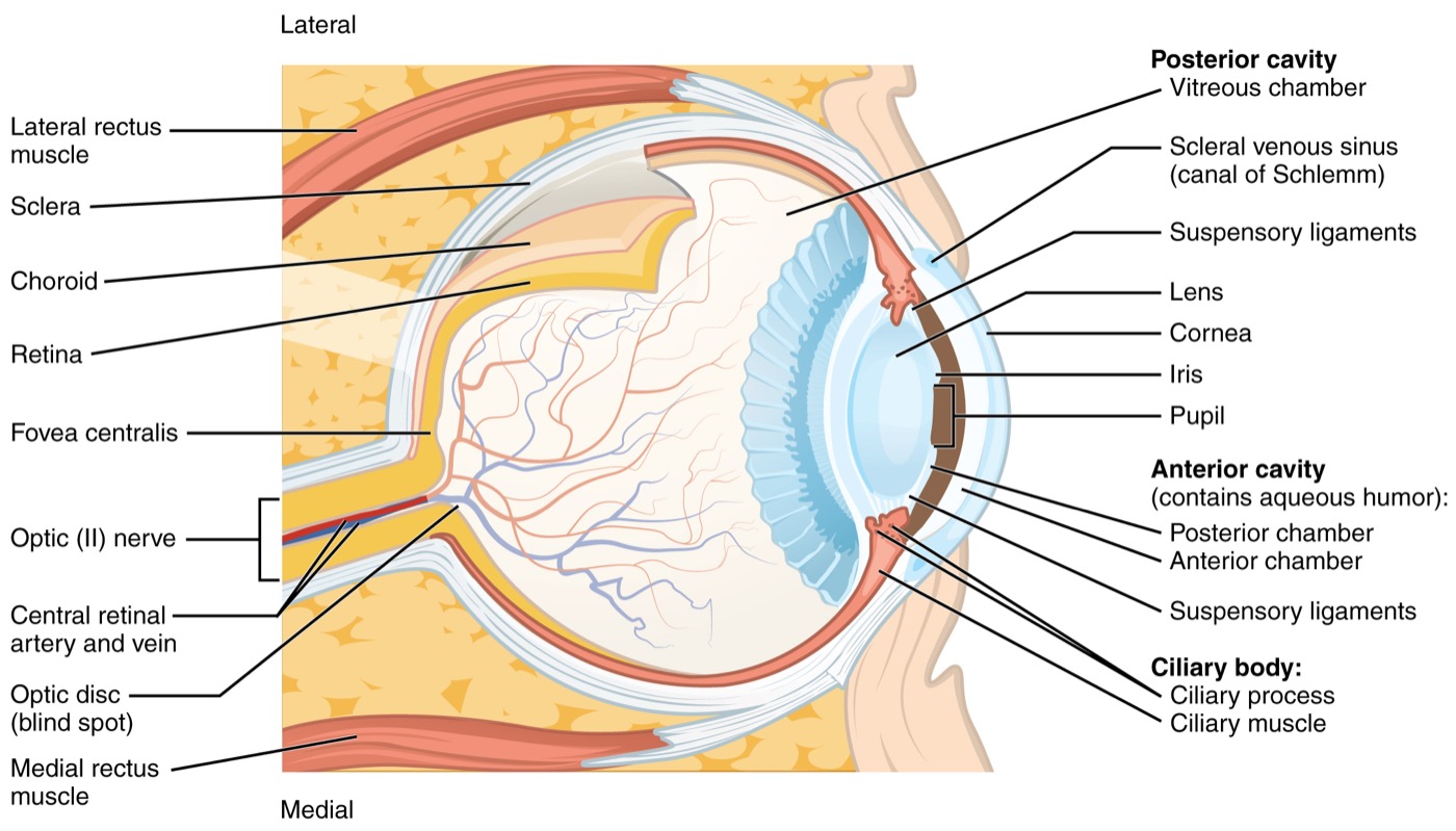
The general senses of chapter seven use receptors spread through the body. The special senses use dedicated organs in the head, wired directly to the brain by cranial nerves rather than through the spinal cord. Vision is the eye and cranial nerve two. Hearing and balance are both the inner ear and nerve eight. Smell is nerve one, and taste uses nerves seven, nine and ten. A useful example of a loop: touch the cornea and both eyes blink, sensation in on nerve five and command out on nerve seven. Someone with facial nerve weakness may be unable to close one eye, so the cornea dries and is damaged without any loss of sensation. Protection depends on the whole loop. The eye About 24 mm long, three layers Cornea: clear, no blood vessels, densely innervated Iris sets the pupil between about 2 and 8 mm Retina lines the back; leaves as the optic nerve Intraocular pressure about 10 to 21 mmHg — glaucoma when drainage fails The eyeball is roughly a sphere about twenty-four millimetres long, built in three layers. The tough outer layer is the white sclera, continuous at the front with the clear cornea, which has no blood vessels and is one of the most densely innervated tissues in the body. The middle layer supplies blood, makes the eye's fluid, holds the lens, and includes the iris, which sets the pupil between about two and eight millimetres. The inner layer is the retina. The fluid in front of the lens is constantly made and drained, and the balance sets the pressure inside the eye, typically about ten to twenty-one millimetres of mercury. When drainage fails, pressure rises and slowly damages the optic nerve: glaucoma, usually silent until the loss is substantial. The light path The cornea does about two thirds of the focusing; the lens is the adjustable part, and it stiffens with age
Two things about the light path surprise engineers. First, the cornea does most of the focusing, not the lens: about forty-three of the eye's roughly sixty dioptres, because refraction is strongest where the change in refractive index is biggest, at the air-cornea surface. That is why reshaping the cornea changes vision so much, and why vision under water is so poor. Second, the lens is the adjustable part, and it stiffens with age. Its focusing range falls from about ten dioptres at twenty to about one by sixty. That is presbyopia, and it is universal: the near point moves from about ten centimetres to about a metre, which is why small print on a device defeats a large share of its users. The retina, and why the pattern of loss matters Receptors Rods: about 90 to 120 million; dim light, no colour Cones: about 6 million; colour; packed in the fovea Only the central couple of degrees is sharp Dark adaptation takes 20 to 30 minutes Low vision is not one thing Cataract: blur, glare, low contrast Glaucoma: periphery lost first Macular degeneration: centre lost Diabetic retinopathy: patchy, fluctuating Something the user is not looking at is something they may not see.
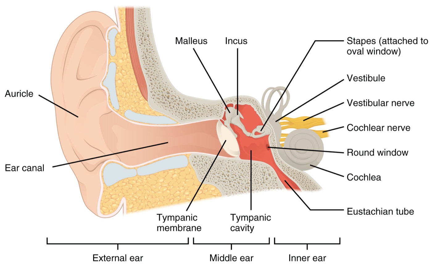
The retina has rods, about ninety to a hundred and twenty million, sensitive in dim light but colour-blind, and cones, about six million, which give colour and are packed into the central fovea. Only the central couple of degrees of your visual field is sharp; the eyes move several times a second and the brain stitches the result. So something the user is not looking at is something they may not see. Low vision is not one condition, and the pattern matters. Cataract brings blur, glare and low contrast. Glaucoma removes the periphery first, so do not put critical information at the edges. Macular degeneration removes the centre. Diabetic retinopathy is patchy and fluctuating. And contrast sensitivity falls with age even when acuity is normal. The ear — three parts, three media Outer: auricle and a 25 mm S-shaped canal to the eardrum Middle: air, three ossicles, the Eustachian tube Inner: fluid — the cochlea and the balance organs The bony inner canal is thin-skinned and easily injured The ear has three parts working in three media. The outer ear is the auricle and an S-shaped canal about twenty-five millimetres long and seven across, ending at the eardrum, which is about a tenth of a millimetre thick. The inner part of the canal is bone covered with very thin skin, which is why an otoscope speculum or an earmould is shaped and inserted with care. The middle ear is air-filled and holds the three smallest bones in the body, the ossicles, with the Eustachian tube equalising pressure to the back of the nose. The inner ear is fluid-filled and contains the cochlea for hearing and the vestibular apparatus for balance. The sound path and the cochlea Air, then mechanical, then fluid — the cochlea sorts frequency by place, and hair cells do not regenerate
Sound travels through air in the canal, becomes mechanical motion in the eardrum and ossicles, and becomes a fluid wave in the cochlea. The cochlea's membrane is narrow and stiff at the base and wide and floppy at the apex, so each frequency peaks at a different place: high frequencies near the base, low near the apex. It is a mechanical spectrum analyser, which is why a cochlear implant stimulates different frequency bands at different depths. About three and a half thousand inner hair cells do most of the signalling, and about twelve thousand outer hair cells act as amplifiers. They even emit faint sounds, which newborn screening detects. And human hair cells do not regenerate: noise, some drugs and age destroy them permanently. Why an older user cannot hear your beep "I can hear you, but I can't understand you, especially in a noisy room." High frequencies are lost first — where consonants live, and where many device beeps sit
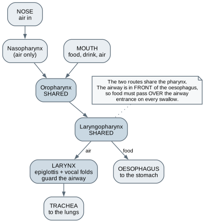
Both noise-induced and age-related hearing loss affect high frequencies first, and that is where the consonants that distinguish words live. So the typical complaint is not I cannot hear, but I can hear you and I cannot understand you, especially in noise. For a designer, a device beep at three or four kilohertz may be exactly the sound an older user cannot hear, while a lower-pitched tone at the same level is perfectly audible. Hearing loss also comes in two kinds. Conductive loss means sound does not reach the cochlea efficiently, from wax, fluid or damaged ossicles, and is often treatable. Sensorineural loss means the cochlea or nerve is damaged; amplification helps audibility but not clarity. Balance — an inertial measurement unit Three semicircular canals: rotation Two otolith organs: linear acceleration and gravity Fused with vision and proprioception The vestibulo-ocular reflex keeps the image still Sensory conflict causes motion sickness — VR creates exactly that The balance organs are effectively an inertial measurement unit. Three semicircular canals at roughly right angles detect rotation of the head, and two otolith organs detect linear acceleration and gravity: they tell you which way is down. The brain fuses that with vision and proprioception, and a reflex counter-rotates the eyes when your head turns, which is why you can read while shaking your head but not while shaking the page. When the senses disagree, the result is motion sickness. A virtual reality headset is a device for creating exactly that disagreement. If you build simulation in VR, latency, frame rate and artificial locomotion matter, and sickness is a design requirement, not a user weakness. The nose and sinuses Turbinates warm, humidify, and filter air in a few centimetres The nose is about half of airway resistance Smell: a few square centimetres; provides most of flavour Little's area on the septum: most nosebleeds Cannula prongs and tube tape cause mucosal pressure injury Inside the nose, scroll-shaped turbinates covered with a richly supplied lining multiply surface area and make the airflow turbulent, so inspired air is warmed, humidified and filtered within a few centimetres. The nose is also the largest single resistance in the airway, about half the total in quiet breathing, which is why it is the narrowest point a nasal cannula, swab or feeding tube must pass. Smell comes from a small patch at the roof of the nasal cavity and provides most of what we call flavour. Its loss, common with age and after viral infection, removes a warning channel for smoke and gas. And pressure from a cannula prong or its tape on the septum can cause a pressure injury within days, on a mucous membrane the skin staging systems do not describe. The crossing Pharynx: shared by air and food Larynx: epiglottis and vocal folds guard the airway The airway is in front of the oesophagus Food passes over the airway on every swallow The pharynx is a muscular tube from the back of the nose to the top of the oesophagus. The upper part carries only air; the lower two parts carry both air and food. The airway lies in front of the oesophagus, so food must pass over the airway entrance on every swallow. The larynx guards it. Its cartilages include the thyroid cartilage at the front of your neck and the cricoid, the only complete ring in the airway. The epiglottis tilts over the entrance during swallowing, and the vocal folds can close the airway completely. The gap between the folds is the narrowest part of the adult airway, and everything in airway management is organised around getting past it. The voice is a source–filter system Source: vibrating vocal folds Fundamental about 85 to 155 Hz in men, 165 to 255 Hz in women Filter: throat, mouth, and nose shape the formants Speech synthesis, recognition, and the electrolarynx use this model If you have done audio work, the voice will be familiar: it is a source-filter system. The vibrating vocal folds are the source, a buzz with a fundamental frequency typically around eighty-five to a hundred and fifty-five hertz in adult men and a hundred and sixty-five to two hundred and fifty-five in adult women, rich in harmonics. The throat, mouth and nose are the filter; their changing shape creates resonances called formants, which make vowels distinguishable. Speech synthesis, speech recognition, and the electrolarynx used after the larynx is removed are all built on this model. Swallowing, and silent aspiration Under a second to protect the airway; silent aspiration happens with no cough at all
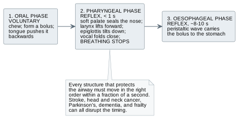
A swallow happens several hundred times a day and is one of the most complex reflex sequences in the body. The oral phase is voluntary. In the pharyngeal phase, in under a second, the soft palate seals the nose, the larynx lifts, the epiglottis tilts, the vocal folds close and breathing stops. Then a wave carries the food down the oesophagus over eight to ten seconds. Difficulty swallowing risks aspiration, material passing below the vocal folds. Normally that triggers a forceful cough, but silent aspiration, with no cough at all, is common after stroke and in other neurological conditions. Absence of a reaction is not evidence of absence of harm. Texture-modified diets are described by the IDDSI framework, eight levels each with a test a carer can do in a kitchen. That is good test design. If it can go in a mouth, assume it can go in an airway Small parts, bitten-off pieces, beads, button cells, magnets — choking and aspiration hazards. A swallowed button cell can burn through tissue in hours. Toy rules treat anything fitting a 31.7 mm cylinder as a small part for under-threes — a convention, not a universal limit
If it can go in a mouth, assume it can go in an airway. Small parts, detachable caps, pieces that can be bitten off a soft simulator, beads, button cells and magnets are choking and aspiration hazards for children, people with dementia, and anyone with a swallowing disorder. Toy regulations use a test cylinder about thirty-two millimetres across for children under three; that is a convention for one population, but it reminds you how large small is. Button cells and magnets are worse than inert parts: a swallowed button cell can burn through tissue in hours. Anything you build with one needs a compartment that cannot be opened without a tool. When the upper airway is bypassed Tracheostomy Tube in the trachea below the larynx Nose no longer humidifies — a heat and moisture exchanger is worn Speech needs a speaking valve Total laryngectomy Trachea opens at a permanent neck stoma No connection between mouth, nose, and lungsOxygen by face mask reaches nothing Two situations change the anatomy in ways you must understand before designing anything used near the neck. A tracheostomy is a tube in the front of the trachea below the larynx. The person breathes through it, so air is no longer warmed and humidified by the nose, which is why they wear a heat and moisture exchanger, and air does not reach the vocal folds unless a one-way speaking valve redirects it. After a total laryngectomy, the larynx is removed and the trachea opens at a permanent stoma in the neck. There is then no connection at all between mouth and nose and the lungs, and oxygen given by face mask reaches nothing. A shower guard, a scarf clip or a training manikin for these users must respect an open airway at the front of the neck. The mouth Enamel: the hardest tissue, about 80 GPa; does not regenerate Tongue: a muscular hydrostat; a few thousand taste buds Saliva about 0.5 to 1.5 litres a day; dry mouth is common Mouth opening about 40 to 55 mm; bite force several hundred newtons Oral hygiene in ventilated patients is infection control In the mouth, enamel is the hardest material in the body, with a stiffness around eighty gigapascals, comparable to aluminium, and like dentine it does not regenerate. The tongue is a muscular hydrostat, muscle with no bone that changes shape at constant volume, carrying most of a few thousand taste buds. The salivary glands make about half a litre to a litre and a half of saliva a day, and dry mouth from medication or radiotherapy makes eating, speaking and wearing a denture hard. A typical adult opens forty to fifty-five millimetres between the incisors, with biting forces of several hundred newtons, numbers that matter for mouthpieces and oral simulators. And oral hygiene in ventilated patients is an infection control measure. The face and neck as a place devices sit Site Why vulnerable Devices that injure it Bridge of the nose Thin skin over bone Masks, goggles, glasses, headsets Behind the ears Thin skin over poorly perfused cartilage Oxygen tubing, straps, glasses arms Nostrils and septum Mucosa; small loaded area Cannula prongs, feeding tubes Lips and mouth Mucosa, moisture, movement Tube holders, bite blocks Front and sides of neck Airway, vessels, nerves near surface Collars, straps, lanyards
Carotid sinus pressure can slow the heart; anything round the neck is a strangulation risk.
These are classic device-related pressure injury sites, and they follow every rule of chapter five: a small area, sustained load, thin tissue over bone or cartilage, moisture, shear, and a user who may not be able to report discomfort. The bridge of the nose suffers under masks and goggles; behind the ears suffers under oxygen tubing; the nostrils under cannula prongs. Health workers in tight respirators for long shifts developed the same injuries on their own faces. The neck adds two hazards: external pressure where the carotid artery divides can slow the heart and drop blood pressure, and anything that encircles the neck is a strangulation risk, which is why clinical lanyards have breakaway clasps. Engineering bridge: optics, acoustics, and airflow Check a label or an alarm against human limits before you build it
Now the engineering bridge: three small pieces of physics that let you check your designs against human limits on paper. Visual angle and legibility Viewing distance Threshold, 6/6 Threshold, 6/12 Threshold, 6/18 0.4 m 0.6 mm 1.2 mm 1.7 mm 1 m 1.5 mm 2.9 mm 4.4 mm 3 m 4.4 mm 8.7 mm 13 mm 6 m 8.7 mm 17 mm 26 mm
Threshold letter height ≈ distance × 0.00145. Then add a reserve of two to three times.
Legibility depends not on size but on the angle a letter subtends at the eye. Standard acuity, called six six or twenty twenty, is reading a letter that subtends five minutes of arc. So the smallest readable letter height is roughly the viewing distance times zero point zero zero one four five. At one metre that is one and a half millimetres; at three metres, about four and a half. For someone with six twelve vision, double it. These are thresholds under test conditions. Comfortable, error-free reading needs two to three times threshold, more in poor light or for someone moving or stressed. That margin is a design decision, and it should be written down. And for lenses: optical power in dioptres is one over the focal length in metres. Decibels Level Typical source 0 dB Threshold of hearing 30 dB Whisper; quiet bedroom 45 to 55 dB Quiet office; many wards by day 60 dB Conversation at 1 m 85 dB Occupational action level for 8 hours 120 to 140 dB Threshold of pain
+6 dB doubles pressure · two equal sources add 3 dB · open space loses 6 dB per doubling of distance
Sound level is logarithmic, in decibels relative to twenty micropascals. A whisper is about thirty, conversation at a metre about sixty, and eighty-five is the occupational action level for an eight hour day. Rules of thumb worth carrying: six decibels more is double the sound pressure, and ten more sounds about twice as loud. Two equal sources add three decibels, not double the number. In open space, level falls about six decibels for each doubling of distance. And exposure trades level against time: under the common three decibel rule, eighty-eight gives you four hours, ninety-one two, and a hundred about fifteen minutes. Workshop machinery reaches those levels easily, and hair cells do not come back. Two pieces of ear engineering Quarter-wave resonator Ear canal: 343 ÷ (4 × 0.025) ≈ 3.4 kHz Boosts 2 to 4 kHz by 10 to 15 dB Block it and you lose the gain; your own voice booms Impedance transformer Eardrum area about 17 times the stapes footplate Ossicular lever about 1.3 Gain ≈ 22 times, about 27 dB Two bits of ear engineering. The ear canal is a tube closed at one end, a quarter-wave resonator. Speed of sound over four times twenty-five millimetres gives about three point four kilohertz, which is why we are most sensitive around two to four kilohertz. Block the canal with an earbud or a hearing aid and you remove that gain, and trap bone-conducted sound so your own voice booms: the occlusion effect. The middle ear is an impedance-matching transformer. Sound going from air into fluid would be almost entirely reflected. The eardrum's effective area is about seventeen times the stapes footplate, and the ossicles add a lever of about one point three, for a pressure gain of about twenty-two, roughly twenty-seven decibels. Lose the ossicles and you lose about that much hearing. Airflow in the upper airway Peak inspiratory flow at rest: about 25 to 30 L/min A nasal cannula supplies about 1 to 6 L/min The rest is room air — the inhaled oxygen fraction is not known from the setting High flows need heating and humidifying In a simulator, the honest output is a range, not a number.
A resting adult's peak inspiratory flow is roughly twenty-five to thirty litres per minute, and much more when breathless. A nasal cannula supplies perhaps one to six. So with every breath the person draws in a mixture of cannula oxygen and room air, in a proportion that depends on how they are breathing. The oxygen fraction actually inhaled is therefore not known from the flow setting; it is estimated, roughly. If you ever build a simulator or training software that displays one, the honest output is a range. Higher flows also dry the nasal lining, which is why high-flow systems heat and humidify the gas. Devices, software, and training technology Clinical devices and software Spectacles, contact lenses, intraocular lenses, tonometers, OCT Otoscopes, audiometers, tympanometers, hearing aids, cochlear implants Cannulas, CPAP masks, laryngoscopes, tracheostomy tubes Magnifiers, simulators, live captioning, Praat for speech Training and maker entry points Airway heads; cricothyroidotomy trainer consumables Otoscopy ears and fundoscopy eyes Cast silicone into machined moulds; real optics; sound rigs Calculation before construction ENT, ophthalmology, audiology, speech and language therapy and dentistry are five separate professions, each with its own equipment. Clinical devices run from spectacles, contact lenses and intraocular lenses to OCT retinal scanners, audiometers and tympanometers, nasal cannulas, CPAP masks, laryngoscopes and tracheostomy tubes. Software includes screen magnifiers, colour-vision simulators, live captioning, and Praat for speech analysis. Good entry points span the workshop: cast and moulded consumables for airway and otoscopy trainers, fundoscopy trainer eyes with a real lens and interchangeable retinal images, electronics for sound and light test rigs, and calculation before construction. Three ordinary materials become hazards here Light sources, sound sources near the ear, and small parts. And nothing you make goes in an eye, ear, nose, mouth, or airway on a person. UV, bright blue LEDs, and lasers damage the eye without pain — photobiological and laser safety standards apply
In this region, three ordinary maker components become hazards. Light: ultraviolet, high-intensity blue LEDs and lasers can damage the eye without pain, so any light source a person might look into must be assessed against the photobiological and laser safety standards, or physically shielded. Sound: a buzzer close to the ear can exceed safe levels even if it seems moderate at arm's length, because the level rises six decibels for every halving of distance. And small parts, as we have seen. Finally, nothing you make goes in an eye, an ear, a nose, a mouth or an airway on a person. Worked example: the label at the back of the room 3 mm
letters on the elbow model — below threshold for everyone beyond about 2 m
At 4 m: 5.8 mm threshold for 6/6, 11.6 mm for 6/12, 23 to 35 mm with a reading reserve
The worked example. Your elbow model's pull to bend label uses letters three millimetres tall, comfortable in the hand. Now the educator wants to use it in front of twenty people, the furthest four metres away. Threshold for standard vision at four metres is about five point eight millimetres; for six twelve vision, about eleven point six; with a reading reserve, twenty-three to thirty-five millimetres. The three millimetre label is below threshold for everyone beyond about two metres. The click fares no better: an invented fifty decibels at half a metre falls to about thirty two at four metres, under a classroom's forty-five to fifty-five. The fix is not a bigger model. It is a large demonstration card, a high-contrast locked flag visible at distance, or a document camera. Then test it from the back of the real room. Workshop exercise and exit criteria Find your blind spot and calculate its angle — about 15 degrees Measure the visual angle of printed words at several sizes Map a room's sound with an uncalibrated phone meter Feel your larynx rise on a swallow Map where glasses or a respirator press on your own face For the workshop: find your own blind spot with a cross and a dot on a card, and calculate its angle; it should be about fifteen degrees. Print a word at five sizes and find the distance at which you can just read each, to see that the angle stays roughly constant. Map a room's sound with a phone meter, recording readings as relative because the phone is uncalibrated, then measure your model's click at three distances. Feel your larynx rise as you swallow, and notice you cannot breathe in at the same moment. And after an hour in glasses or a respirator, map the red marks on your face and name the bone or cartilage under each. What this chapter does not establish You cannot test anyone's vision or hearing You cannot assess a swallow or recommend a diet texture You have not learned airway management Light and sound safety are settled by standards and measurement Five separate specialties — enter each through a named partner Next — Circulation, breathing, and the wider body.
The limits. A letter chart and a phone app show you the principles; testing vision and hearing are clinical measurements with calibrated equipment. Swallowing assessment and diet texture belong to speech and language therapists; IDDSI appears here as good measurement design, not something to apply. The anatomy lets you follow a conversation about airways, not manage one. Light and sound safety are settled by the applicable standard and a measurement, not by whether it seemed fine on the bench. And these are five separate specialties, each entered through a named partner. Next, circulation, breathing and the wider body.