Chapter 9
Circulation, breathing, and the wider body
Comprehension, and restraint — the gap between displaying a number and supporting a decision
Human Biology for the Medical Maker
Why this chapter exists
- Follow a clinical conversation about the heart and lungs
- Recognise when a small change touches an essential function
- "Make the strap a bit tighter" — does it cross a superficial artery?
- A monitor display is easy to make
Displaying a number and supporting a clinical decision are separated by the most important boundary in this book.
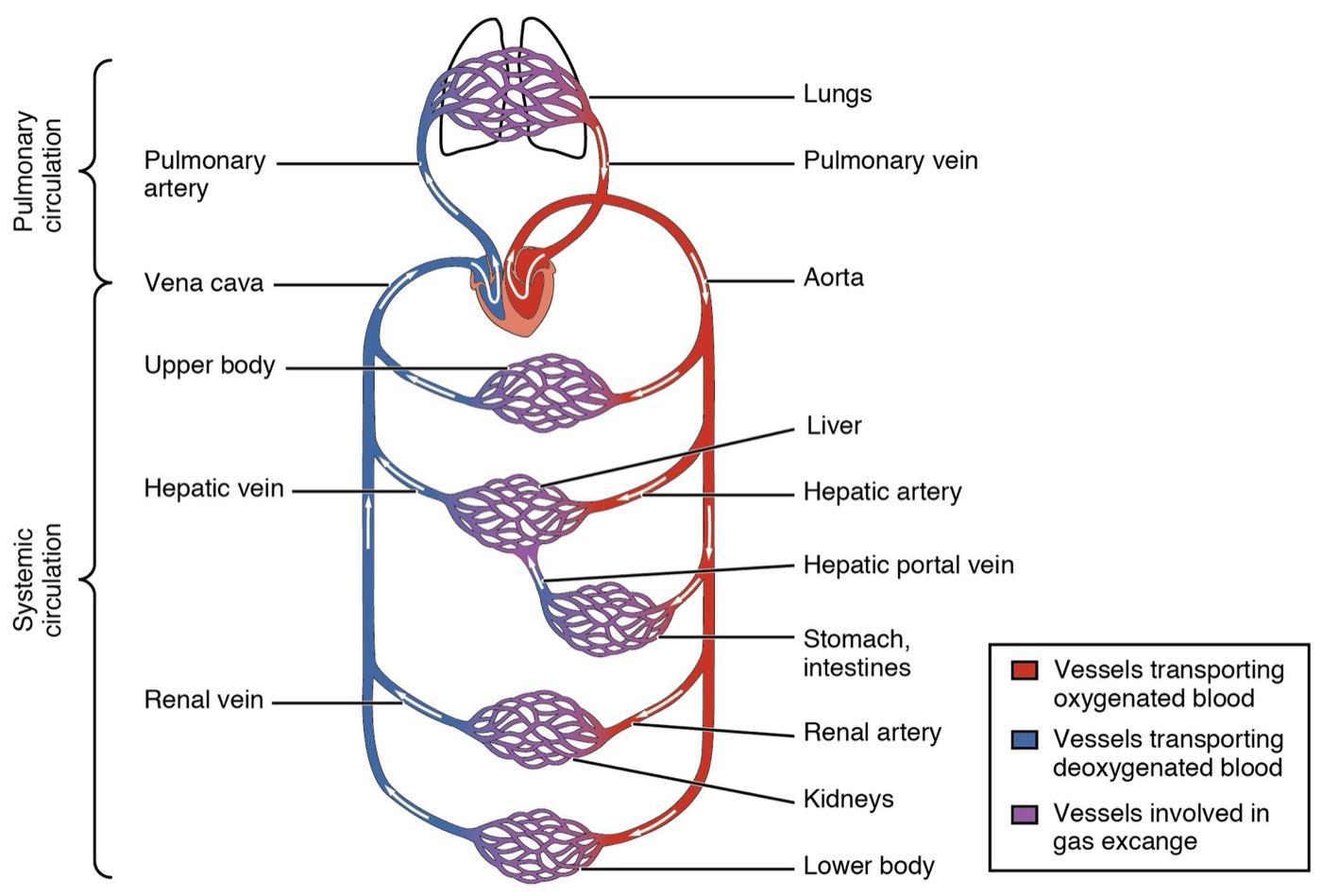
The heart and the two circuits
- Right heart pumps to the lungs; left heart to the body
- Body, venae cavae, right atrium, tricuspid, right ventricle
- Pulmonary arteries, lungs, pulmonary veins, left atrium
- Mitral valve, left ventricle, aortic valve, aorta, body
Arteries and veins
An artery carries blood away from the heart; a vein carries it toward the heart. That is the whole definition.
The pulmonary arteries carry oxygen-poor blood. "Arteries are red, veins are blue" is wrong for the lungs.
Vessel types
| Vessel | Function | Mechanical note |
|---|
| Artery | High-pressure delivery | Elastic recoil smooths flow — a Windkessel |
| Arteriole | Regulates flow into capillary beds | The main site of resistance control |
| Capillary | Exchange of gas, nutrients, waste | 5 to 10 micrometres; one cell thick |
| Vein | Low-pressure return; holds most blood | Valves; muscle contraction pumps |
| Lymphatic | Returns tissue fluid | Failure causes lymphoedema |
The cardiac cycle in numbers
| Quantity | Typical resting adult | Relationship |
|---|
| Heart rate | 60 to 80 per minute | — |
| Stroke volume | about 70 mL | Ejected per beat |
| Cardiac output | about 5 L/min | CO = HR × SV |
| Ejection fraction | 55 to 70% | Stroke volume over filling volume |
| Blood volume | about 5 L | Circulates about once a minute |
"Lub" is the AV valves closing; "dub" is the outflow valves closing.
The conduction system and the ECG
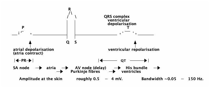
P — atria · QRS — ventricles · T — ventricular recovery. Reading a trace is a clinical skill
Engineering bridge: haemodynamics
Productive analogies — if you know where they break
Ohm's law and Poiseuille's law
The analogues
- Pressure drop = flow × resistance
- Q = π ΔP r⁴ ÷ 8ηL
- Halve the radius: one sixteenth the flow
- Arterioles are the flow-control valves
Where they fail
- Blood is not Newtonian
- Vessels are compliant, tapering, branching
- Flow is pulsatile, sometimes turbulent
- The resistor changes value with the flow
Anything pressing on tissue is a flow-control device, intended or not.
Pressures around the circuit
| Location | Approximate pressure | mmHg |
|---|
| Aorta and large arteries | systolic / diastolic | 120 / 80 |
| Arterioles | falling | 60 to 30 |
| Capillaries | arterial to venous end | 35 to 15 |
| Venae cavae | near zero | 0 to 5 |
| Pulmonary artery | about one fifth of systemic | 25 / 10 |
An interface pressure of 20 kPa is 150 mmHg — several times capillary pressure.
Breathing
Ventilation, mechanics, and gas exchange
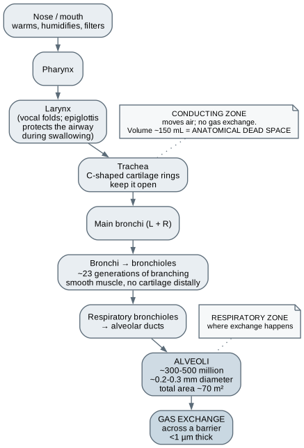
The airway and dead space
- Conducting zone moves air; no exchange
- About 150 mL of anatomical dead space
- Tidal 500 mL: 350 mL useful
- Half the volume, double the rate: 100 mL useful
Mechanics of ventilation
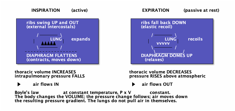
The body changes the volume; the pressure follows; the lungs do not inflate themselves
Three properties an engineer recognises
| Property | Definition | Clinical relevance |
|---|
| Compliance | Volume change per pressure change | Stiff lungs need more pressure; lost recoil traps air |
| Airway resistance | Pressure drop per unit flow | Asthma narrows airways — radius to the fourth again |
| Surfactant | Lowers surface tension, more so in small alveoli | Stabilises alveoli; absent in premature infants |
Lung volumes, typical adult
| Volume | Typical | Meaning |
|---|
| Tidal volume | 500 mL | One quiet breath |
| Respiratory rate | 12 to 20 per minute | At rest |
| Minute ventilation | about 6 L/min | Tidal volume times rate |
| Vital capacity | about 4600 mL | Maximum breath out after maximum in |
| Residual volume | about 1200 mL | Cannot be exhaled |
| Total lung capacity | about 5800 mL | Vital capacity plus residual |
Spirometry compares a person with predicted values — a measurement-design problem in itself.
Gas exchange
Three processes, matched
- Ventilation: air to the alveoli
- Diffusion: across the barrier
- Perfusion: blood past the alveoli
- V/Q mismatch: shunt or dead space
Haemoglobin
- Alveolar oxygen about 104 mmHg; venous blood arrives at 40
- Most oxygen rides on haemoglobin
- Sigmoid curve: loads in the lungs, unloads in tissue
- Shifts right with heat, CO2, and acid
What the vital signs actually measure
| Sign | Obtained by | What it is not |
|---|
| Heart rate | Palpation, ECG, PPG | Not cardiac output or perfusion |
| Blood pressure | Cuff or arterial line | Not flow — pressure can hold while flow fails |
| Respiratory rate | Observation or impedance | Not tidal volume or gas exchange |
| SpO2 | Pulse oximeter | Not ventilation, CO2, or oxygen delivery |
| Temperature | Many sites, all different | Not interchangeable between sites |
| Pain score | Self-report | Not a physical measurement — it is the person's |
How a pulse oximeter works
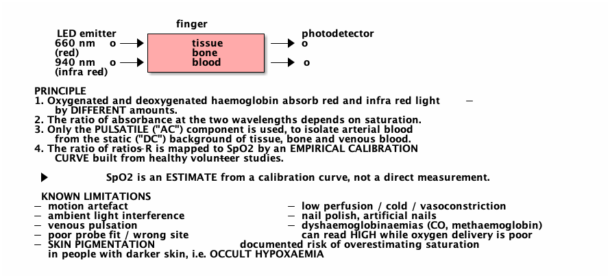
SpO2 is an estimate from an empirical calibration curve, not a direct measurement
The most important lesson for anyone who writes software
Pulse oximeters overestimate saturation more often in people with darker skin — true low oxygen can be missed.
A device calibrated on a non-representative population performs worse on the people it was not calibrated on.
The four layers: phenomenon, sensor, display, interpretation
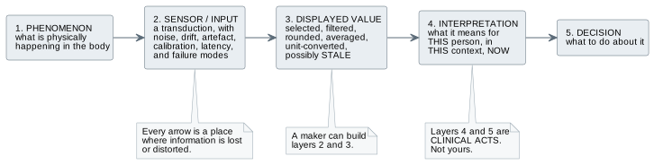
A maker can build layers 2 and 3. Layers 4 and 5 are clinical acts.
Rules for any physiological display — even a mock-up
- Simulation status unmissable; units beside every number
- Show the age of a value; never repeat a stale one as current
- Distinguish no data from zero
- Make invalid input explicit; never clamp silently
- Paused visibly different from live; handle interruption
- Do not label anything "normal"
The other systems: a map
| System | Core function | A maker might meet it as |
|---|
| Renal and urinary | Fluid, electrolytes, pH, waste | Catheter care, fluid measurement, continence aids |
| Digestive | Breakdown, absorption | Feeding aids, enteral connectors, stomas |
| Endocrine | Hormonal regulation | Glucose monitoring, insulin delivery |
| Immune and lymphatic | Defence, fluid return | Infection control, lymphoedema garments |
| Reproductive | Reproduction and hormones | Obstetric and examination trainers |
Fluid compartments and thermoregulation cut across all of them — an enclosure is a thermal decision.
Devices, software, and training technology
Clinical devices — mostly not for you
- ECG, oximeters, BP monitors, capnographs
- Defibrillators, pacemakers, ventilators, CPAP
- Infusion pumps, ECMO, central monitoring
- Alarm fatigue: the dominant human factors problem
Software and training
- Early warning scores; PhysioNet, WFDB, NeuroKit2
- CPR manikins, AED trainers, patient simulators
- Auscultation and ECG rhythm simulators; test lungs
- Moulage — casting, fabrics, dressings
The mechanical test lung — an unusually good project
- A bellows with a settable spring (compliance) and orifice (resistance)
- Sewn or moulded bellows, turned orifice plates, machined end plates
- Springs selected by measured rate; no electronics needed
- Add a pressure sensor later — outside the gas path
Change the spring and the pressure changes; narrow the orifice and resistance rises as radius to the fourth.
Worked example: the classroom mock monitor
Build it with
- A permanent "SIMULATION ONLY" banner
- Obviously fictional data; units everywhere
- Live, paused, and stale visibly different
- A visible version number
Do not
- Connect it to any real device or system
- Use real patient data
- Invent alarms, thresholds, or interpretation
- Deploy it where it could be read as real
Almost none of this is programming. The engineering is in the framing.
Workshop exercise and exit criteria
- Draw both circuits from memory, marking where exchange happens
- Draw the air path; compare alveolar ventilation at 500 and 250 mL
- Calculate MAP for 118/76, cardiac output for 72 × 68 mL, a 20% radius drop
- Build the mock monitor with one misleading behaviour — then fix it
- Trace a strap you own across a superficial artery or nerve; ask, don't experiment
What this chapter does not establish
- You cannot interpret any measurement
- You cannot set alarm limits or thresholds
- You cannot build a monitoring device
- Demonstrating mechanics does not demonstrate physiology
- The numbers are orientation, not reference ranges
Next — Kidneys, gut, and hormones: the internal regulators.